


Knee & Lower Leg
Conditions
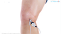
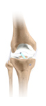
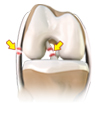
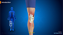
Knee Anatomy
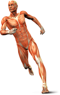
The knee is a complex joint made up of different structures including bones, tendons, ligaments and muscles. They all work together to maintain normal function and provide stability to the knee during movement.
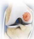
Articular Cartilage Injury
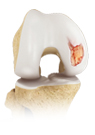
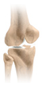
Articular or hyaline cartilage is the tissue lining the surface of the two bones in the knee joint. Cartilage helps the bones move smoothly against each other and can withstand the weight of the body during activities such as running and jumping. Articular cartilage does not have a direct blood supply to it so has less capacity to repair itself. Once the cartilage is torn it will not heal easily and can lead to degeneration of the articular surface, leading to development of osteoarthritis.
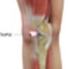
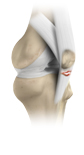
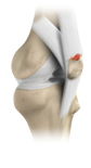
Jumper's Knee
Patellar tendinitis, also known as "jumper's knee" is an inflammation of the patellar tendon that connects your kneecap (patella) to your shinbone. This tendon helps in extension of the lower leg. Patellar tendinitis usually results from repetitive trauma or overuse, particularly from sports activities involving jumping such as basketball or volleyball. Therefore, this condition is also known as jumper's knee. Rarely, this condition may also occur because of an acute injury to the tendon that has not healed properly.
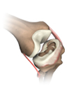
Knee Anterior Cruciate Ligament Injury
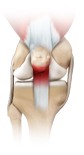
The anterior cruciate ligament, or ACL, is one of the major ligaments of the knee that is located in the middle of the knee and runs from the femur (thigh bone) to the tibia (shin bone). It prevents the tibia from sliding out in front of the femur. Together with posterior cruciate ligament (PCL) it provides rotational stability to the knee.
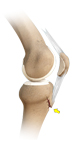
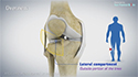
Knee Lateral Collateral Ligament Injury
Lateral collateral ligament (LCL) is a thin set of tissues present on the outer side of the knee, connecting the thighbone (femur) to the fibula (side bone of lower leg). It provides stability as well as limits the sidewise rotation of the knee. Tear or injury of LCL may cause instability of the knee that can be either reconstructed or repaired to regain the strength and movement of the knee.
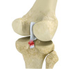
Knee Medial Collateral Ligament Injury
The medial collateral ligament (MCL) is the ligament that is located on the inner part of the knee joint. It runs from the femur (thighbone) to the top of the tibia (shinbone) and helps in stabilizing the knee. Medial collateral ligament (MCL) injury can result in a stretch, partial tear, or complete tear of the ligament. Injuries to the MCL commonly occur as a result of a pressure or stress on the outside part of the knee. Anterior cruciate ligament (ACL) may be torn along with a MCL injury.
Knee Pain

Osgood-Schlatter disease refers to a condition of an overuse injury that occurs in the knee region of growing children and adolescents. This is caused by inflammation of the tendon located below the knee cap (patellar tendon). Children and adolescents who participate in sports such as soccer, gymnastics, basketball and distance running are at higher risk of this disease.
Kneecap Fractures
Coming Soon
Kneecap Bursitis
AA bursa is a small fluid-filled sac found between soft tissues and bones. It lubricates and acts as a cushion to decrease friction between bones when they move. Bursitis refers to the inflammation and swelling of the bursa. Inflammation of the bursa in front of the kneecap (patella) is known as kneecap bursitis or prepatellar bursitis.
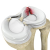
Meniscus Tears

Meniscus tear is the commonest knee injury in athletes, especially those involved in contact sports. A suddenly bend or twist in your knee cause the meniscus to tear. This is a traumatic meniscus tear. Elderly people are more prone to degenerative meniscal tears as the cartilage wears out and weakens with age. The two wedge-shape cartilage pieces present between the thighbone and the shinbone are called meniscus. They stabilize the knee joint and act as “shock absorbers”.
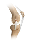
Multi-Ligament Injury
Ligaments are the fibrous tissue brands connecting the bones in the joint and stabilizing the joint. Knee joint has 2 sets of ligaments-collateral ligaments (medial and collateral ligaments) that connect the bones on outer side of the knee and cruciate ligaments (anterior cruciate ligament and posterior cruciate ligament) those present inside the joint. Multi-ligament injury is the injury to multiple ligaments at the same time. Damage to three or more ligaments may cause joint dislocation.
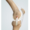
Patellar Tendon Injuries & Tears
Patellar tendon rupture is the rupture of the tendon that connects the patella (knee cap) to the top portion of the tibia (shin bone). The patellar tendon works together with the quadriceps muscle and the quadriceps tendon to allow your knee to straighten out.
Pediatric Tibial Tubercle Fractures
Tibial tubercle fractures are quite rare occurrences that typically affect physically active adolescents between the age 14 and 17. It is caused from violent tensile forces exerted over the tibial tuberosity (a bulge in the tibial bone) during activities involving sudden contraction of the knee extensors (springing and jumping). A history of Osgood-Schlatter disease in the family may increase susceptibility to tibial tubercle fracture.
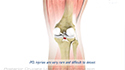
Posterior Cruciate Ligament Injury
PCL injuries are very rare and are difficult to detect than other knee ligament injuries. Cartilage injuries, bone bruises, and ligament injuries often occur in combination with PCL injuries. Injuries to the PCL can be graded as I, II or III depending on the severity of injury. In grade I the ligament is mildly damaged and slightly stretched, but the knee joint is stable. In grade II there is partial tear of the ligament. In grade III there is complete tear of the ligament and the ligament is divided into two halves making the knee joint unstable.
Quadriceps Tendon Injuries & Tears
Quadriceps tendon is a thick tissue located at the top of the kneecap. The quadriceps tendon works together with the quadriceps muscles to allow us to straighten our leg. The quadriceps muscles are the muscles located in front of the thigh.
Runner's Knee
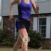
Runner's knee, also called patellofemoral pain syndrome refers to pain under and around your kneecap. Runner’s knee includes several medical conditions such as anterior knee pain syndrome, patellofemoral malalignment, and chondromalacia patella that cause pain around the front of the knee. As the name suggests, runner’s knee is a common complaint among runners, jumpers, and other athletes such as skiers, cyclists, and soccer players.
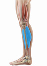
Shin Splints
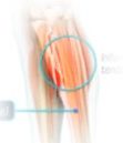
“Shin splints” is used to describe the pain and inflammation of the tendons, muscles and bone tissue around the tibia or shine bone (a large bone in the lower leg). It occurs because of vigorous physical activity such as exercise or sports. The condition is also referred to as medial tibial stress syndrome (MTSS).
Stress Fractures
Coming soon
Tennis Leg – Calf Injuries
Coming soon
Knee Arthritis
Arthritis is a general term covering numerous conditions where the joint surface or cartilage wears out. The joint surface is covered by a smooth articular surface that allows pain free movement in the joint. This surface can wear out for a number of reasons; often the definite cause is not known. When the articular cartilage wears out the bone ends rub on one another and cause pain. This condition is referred to as Osteoarthritis or "wear and tear" arthritis as it occurs with aging and use. It is the most common type of arthritis.
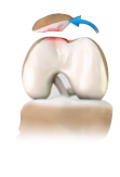
Osteochondral Autograft
Osteochondral autografting (OCG) is a surgery to repair damaged articular cartilage that lines the ends of bones in a joint. An osteochondral autograft is a piece of tissue taken from a healthy section of the joint and transplanted to replace the chondral defects in the joint. There are two techniques used in osteochondral autografting: mosaicplasty and osteochondral autograft transfer system (OATS).
Osteochondral Allograft
An osteochondral allograft is a piece of tissue taken from a diseased donor to replace damaged cartilage that lines the ends of bones in a joint. A section of cartilage and bone is removed, shaped to precisely fit the defect and then transplanted to reconstruct the damage.
Patellofemoral Instability
Patella (knee cap) is a protective bone attached to the quadriceps muscles of the thigh by quadriceps tendon. Patella attaches with the femur bone and forms a patellofemoral joint. Patella is protected by a ligament which secures the kneecap from gliding out and is called as medial patellofemoral ligament (MPFL).
Videos

Procedures
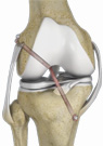
ACL Reconstruction
The anterior cruciate ligament is one of the major stabilizing ligaments in the knee. It is a strong rope like structure located in the centre of the knee running from the femur to the tibia.
Knee Dislocation
Coming soon
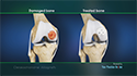
Advanced Cartilage Restoration
Articular or hyaline cartilage is the tissue lining the surface of the two bones in the knee joint. Cartilage helps the bones move smoothly against each other and can withstand the weight of the body during activities such as running and jumping. Articular cartilage does not have a direct blood supply to it so has less capacity to repair itself. Once the cartilage is torn it will not heal easily and can lead to degeneration of the articular surface, leading to development of osteoarthritis.
Joint Preservation and Reconstruction
Coming soon
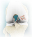
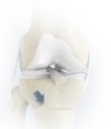
Knee Arthroscopy
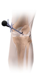
The arthroscope is a fiber-optic telescope that can be inserted into a joint (commonly the knee, shoulder and ankle) to evaluate and treat a number of conditions. A camera is attached to the arthroscope and the picture is visualized on a TV monitor. Most arthroscopic surgery is performed as day surgery and is usually done under general anesthesia. Knee arthroscopy is common, and millions of procedures are performed each year around the world.
Meniscal Repairs
Meniscus is the C-shaped two pieces of cartilage located between thighbone and shin bone that act as shock absorbers and cushion the joints. Meniscus distributes the body weight uniformly across the joint and avoids the pressure on any one part of the joint and development of arthritis. Being the weight bearing part, meniscus is prone to wear and tear and meniscal tear is one of the common knee injuries. Meniscal tear may be developed by people of all ages and is more common in individuals who play contact sports.
Multi-ligament Knee Reconstruction
Ligaments are fibrous tissue bands that connect bones and stabilize joints. The knee joint has four major ligaments – the anterior cruciate ligament, posterior cruciate ligament, lateral collateral ligament, and medial collateral ligament.
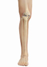
High Tibial Osteotomy
High tibial osteotomy is commonly used for patients with osteoarthritis that is isolated to a single compartment (unicompartmental osteoarthritis). It is also performed for treating a variety of knee conditions such as gonarthrosis with varus or valgus malalignment, osteochondritis dissecans, osteonecrosis, posterolateral instability, and chondral resurfacing.
Exertional Compartment Syndrome Surgery
Exertional compartment syndrome, also called chronic compartment syndrome, is a condition that causes pain or cramps in the legs during exercise. This pain usually lessens on stopping the activity. It most often occurs in the front compartment of the lower leg. Athletes participating in sports such as running, biking, or swimming which require repetitive movements are at a greater risk of developing this condition.