


Cartilage Repair
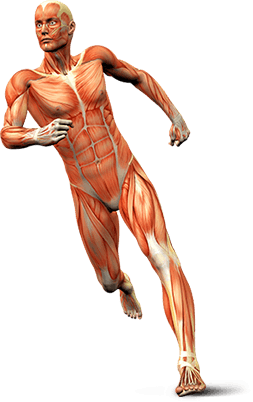
The hip joint is the largest weight-bearing joint in the body. It is a ball and socket joint where the head of the femur (thigh bone) is the ball and the pelvic bones form a socket called the acetabulum. The bone surfaces in the joint are covered by smooth cartilage which acts as a cushion and enables the bones to easily glide over each other during movement. A rim of cartilage called the labrum is present at the edge of the socket and increases the depth of the socket providing stability to the joint.
Damage to the articular cartilage can cause severe pain in the hip or groin, a locking sensation in the hip joint, significant restriction of hip movement and difficulty walking. This can result from trauma, tissue impingement, loose bodies in the joint, hip dislocation, dysplasia, osteonecrosis, degenerative arthritis and other conditions.
The management of cartilage injuries depends on the severity of the injury and includes non-surgical and surgical modalities of treatment. Non-surgical treatment includes a healthy diet, regular exercise and avoidance of aggravating activities. Surgery is performed to treat cartilage injuries when non-surgical options are ineffective and pain persists.
Hip cartilage repair is usually performed by arthroscopy, also referred to as keyhole surgery or minimally invasive surgery. This involves the use of an arthroscope, a narrow tube with a tiny camera attached on the end, to assess the damage to the hip cartilage. Your surgeon makes 2 to 3 small incisions around the hip joint. The arthroscope is inserted through one of the incisions and the camera attached to the arthroscope helps visualize the joint on a monitor. A sterile solution is pumped into the joint to clear the view and increase the space for surgery. Specially designed instruments are inserted through the other incisions. During the surgery, any loose fragments, small pieces of bone and cartilage floating within the joint are removed. Areas of excess bone growth are trimmed. A torn labrum may be repaired depending on the extent of the injury. After the completion of the procedure, the arthroscope is removed and incisions are closed.
Other arthroscopic surgeries that may be recommended to manage cartilage injuries include:
Microfracture surgery: This technique to treat cartilage injuries involves stimulating the formation of new articular cartilage by drilling numerous tiny holes in the bone underneath the damaged cartilage. This results in the formation of blood clots within the damaged cartilage, which stimulates the growth of new cartilage known as fibrocartilage. Although, the fibrocartilage formed is different from the normal hyaline cartilage, it can provide significant improvement in the symptoms.
Autologous Cartilage Implantation (ACI): In this procedure, the cartilage, harvested from the patient or a deceased donor, is cultured and later implanted over the damaged area of the joint.
Matrix-induced Autologous Chondrocyte Implantation (MACI): This technique employs cultured chondrocytes (the cells which produce the cartilage) to repair the articular cartilage damage. These chondrocytes are inserted onto a layer of collagen which is then implanted over the damaged area of the joint.