


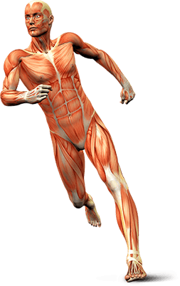


Rotator Cuff Tear
Rotator cuff is the group of tendons in the shoulder joint providing support and enabling wider range of motion. Major injury to these tendons may result in tear of these tendons and the condition is called as rotator cuff tear. It is one of the most common causes of shoulder pain in middle aged adults and older individuals. It may occur with repeated use of arm for over head activities, while playing sports or during motor accidents. Rotator cuff tear causes severe pain, weakness of the arm, and crackling sensation on moving shoulder in certain positions. There may be stiffness, swelling, loss of movements, and tenderness in the front of the shoulder.
Rotator cuff tear is best viewed on magnetic resonance imaging. Symptomatic relief may be obtained with conservative treatments – rest, shoulder sling, pain medications, steroidal injections and certain exercises. However surgery is required to fix the tendon back to the shoulder bone. Rotator cuff repair may be performed by open surgery or arthroscopic procedure. In arthroscopy procedure space for rotator cuff tendons will be increased and the cuff tear is repaired using suture anchors. These anchor sutures help in attaching the tendons to the shoulder bone. Following the surgery you may be advised to practice motion and strengthening exercises.
Arthroscopic Rotator Cuff Repair
The rotator cuff refers to a group of four tendons that attach four shoulder muscles to the upper arm (humerus) and hold it in the shoulder joint. Many shoulder problems are caused by injuries to the rotator cuff.
Surgery to repair the rotator cuff has traditionally been done through a large shoulder incision, about 6-10cm long, and the muscle over the rotator cuff was separated. Newer, advanced surgical techniques have been developed to minimize pain and recovery time. Arthroscopic rotator cuff repair is a minimally invasive surgery performed through tiny incisions, about 1 cm each, with an arthroscope.
The arthroscope is a small fiber-optic viewing instrument made up of a tiny lens, light source and video camera. The surgical instruments used in arthroscopic surgery are very small (only 3 or 4 mm in diameter) but appear much larger when viewed through an arthroscope.
The television camera attached to the arthroscope displays the image of the joint on a television screen, allowing the surgeon to look throughout the shoulder-at cartilage, ligaments, and the rotator cuff. The surgeon can determine the amount or type of injury, and then repair or correct the problem.
The benefits of arthroscopy compared to the alternative, open shoulder surgery, include:
- Smaller incisions
- Minimal soft tissue trauma
- Less pain
- Faster healing time
- Lower infection rate
- Less scarring
- Earlier mobilization
- Usually performed as outpatient day surgery
Massive Irreparable Tear - Xenograft
A massive irreparable rotator cuff tear is the tear of size > 5 cm in diameter and involves more than one tendon. It is usually found out during a diagnostic scan or at surgery. It occurs more commonly in older people and is unusual in individuals below the age of 60 years. Large irreparable tears happen in patients with rotator cuff degeneration and atrophy of the rotator cuff muscle and the cause for this is attributed to genetic changes or age or a combination of both.
Treatment of these massive degenerative tears with direct tendon repair has encountered very high rates of failure. Large tears that can be reattached and fixed back to the bone have shown unacceptable rates of nonhealing. Issues such as inadequate healing and poor tissue quality have led to the use of patch grafts to bridge the difficult tears. A patient who has a larger tear that can be repaired back to the bone, but bears a higher probability of re-tear will be considered for augmentation procedure with a graft.
Xenografts
Xenografts are the tissue grafts obtained from other species and the procedure is called the xenotransplantation. The xenografts for massive irreparable rotator cuff tear may be obtained from different species and some of them include:
- Porcine small intestine submucosa: Porcine small intestine submucosa material (obtained from pigs) has been recommended for usage in rotator cuff augmentation. These materials are made up of 10-layer laminates of porcine collagen that are processed and sterilized so that it is free from cellular or immunologic DNA components. Because of the high risks and low benefits associated with the use of this material, a newer 8-layer lightly crosslinked material which is sterilized using gamma irradiation is marketed for rotator cuff use. This material seems to have lower pull-out strength compared to other augmentation materials
- Bovine dermis: Fetal bovine dermis, which is a single layer of bovine dermal xenograft that is processed to take away the cells, fats and carbohydrates from it and then sterilized with ethylene dioxide, may be used as a graft
- Porcine dermal collagen: Porcine dermal collagen is crosslinked and is sterilized with gamma irradiation but has mixed results in clinical use
- Equine pericardium: Equine pericardium is the most recently marketed collagenous xenograft. This is a crosslinked, decellularized and sterilized product which has been approved by the FDA for its use in the repair of soft tissue defects and tissue reinforcement as well as tendon repair procedures. However, there are no considerable clinical reports of use of this material in rotator cuff
Click on the topics below to find out more from the Orthopaedic connection website of American Academy of Orthopaedic Surgeons.